

A new lump, persistent abdominal pain, urinary change, jaundice, or a swollen leg can justify diagnostic imaging, but the correct scan and timing depend on the symptom. By the end, you will know when to seek emergency care, which ultrasound examination fits your concern, how to prepare, and when another test or clinical review is needed.
Key takeaways
- Seek urgent care for severe pain, fainting, heavy bleeding, or sudden weakness.
- Match the scan to the clinical question instead of booking a whole-body ultrasound.
- Follow fasting and bladder instructions for clearer images and fewer delays.
- Ask how the report will answer your clinician’s specific diagnostic question.
Recognise when symptoms call for ultrasound—and when they need urgent care
Persistent right-upper-quadrant pain, suspected gallstones, pelvic pain, a new lump, urinary symptoms, jaundice, or suspected kidney obstruction can justify an ultrasound appointment. Ultrasound for abdominal pain is useful when the clinical question involves the gallbladder, liver, kidneys, bladder, or abdominal aorta. Ask a clinician which examination to book rather than choosing a “whole-body” scan.
| Situation | Best next step | Why |
|---|---|---|
| Routine symptoms without danger signs | Book the advised ultrasound | Targeted imaging can assess gallstones, pelvic organs, thyroid nodules, or urinary obstruction. |
| Swollen, warm, painful one-sided leg | Arrange urgent venous Doppler through a clinician or urgent-care service | Waiting several days for a routine appointment could delay treatment for deep-vein thrombosis. |
| Severe abdominal pain with faintness, cold sweat, confusion, marked weakness, or a rigid abdomen | Seek emergency assessment | Shock, bleeding, infection, or another time-critical condition may need treatment beyond ultrasound. |
| Sudden weakness on one side, severe chest pain, breathing difficulty, fainting with heavy bleeding, or a cold, pale painful limb | Call emergency services | These symptoms can signal stroke, heart attack, major bleeding, or loss of circulation. |
Jaundice warrants prompt medical review; arrange a jaundice ultrasound scan appointment only through that clinical pathway. During pregnancy, vaginal bleeding, one-sided pelvic pain, shoulder-tip pain, dizziness, or fainting needs urgent assessment, not a routine scan. A very early normal or inconclusive scan cannot exclude every pregnancy problem.
Match the clinical question to the right ultrasound examination
Choose the examination by the body part and clinical question, not by booking a generic “whole-body ultrasound.” These types of ultrasound examinations are not interchangeable.
| Examination | Best matched question | Key point |
|---|---|---|
| Abdominal ultrasound | Gallstones, right-upper-quadrant pain, jaundice, liver or pancreas concerns | Covers upper-abdominal organs; fasting instructions often apply |
| Pelvic ultrasound | Uterus, ovaries, pelvic pain, abnormal bleeding | Usually performed over the lower abdomen |
| Transvaginal ultrasound | Detailed view of the uterus, ovaries, endometrium, or early pregnancy | Uses an internal probe; ask about transvaginal ultrasound consent and privacy |
| Obstetric ultrasound | Pregnancy location, growth, heartbeat, or anatomy | Timing affects what can be seen; an early inconclusive scan may need review |
| Breast ultrasound | A palpable lump or focal breast symptom | Complements examination and mammography when indicated |
| Thyroid ultrasound | Thyroid nodule, neck swelling, or known abnormality | Do not use indiscriminate screening without clinical assessment |
| Scrotal ultrasound | Testicular pain, swelling, lump, or injury | Sudden severe pain needs urgent assessment |
| Renal ultrasound | Flank pain, urinary symptoms, obstruction, or kidney abnormality | Assesses kidneys and often the bladder |
| Doppler ultrasound | Blood flow, suspected deep-vein thrombosis, or blocked vessels | The protocol must match the vessel and question |
| Musculoskeletal ultrasound | Tendon, muscle, ligament, joint, or soft-tissue lump | Dynamic movement can reveal problems missed at rest |
The referral or request should name the suspected problem, relevant side, and body region. That helps the sonographer obtain the right views and add Doppler when blood-flow information matters.
Ask what preparation applies: an abdominal ultrasound may require fasting, while a pelvic ultrasound may require a full bladder. Follow the center’s instructions rather than applying one rule to every scan.
Prepare correctly for the scan you have been advised to get
Ultrasound preparation depends on the examination, so follow the imaging center’s written instructions rather than fasting or drinking water by default. Fasting before abdominal ultrasound commonly means avoiding food for several hours. This reduces bowel gas and keeps the gallbladder expanded for clearer images; ask whether you may drink water or take regular medicines.
1. For a pelvic ultrasound, arrive with a full bladder for pelvic ultrasound if instructed. Drink the stated amount of water at the stated time and avoid urinating until the first images are complete. The bladder moves bowel away and creates a clearer window to the uterus and ovaries.
Tell staff if the pressure becomes painful.
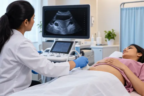
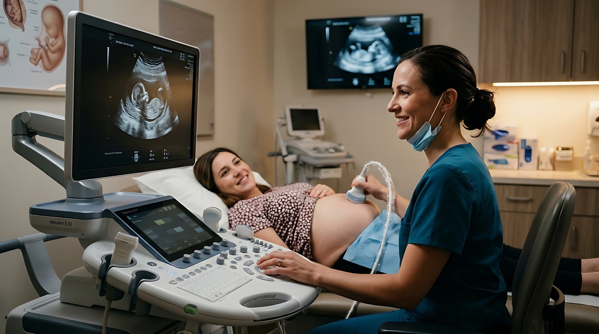
2. During a transabdominal pelvic scan, you lie on your back while gel and a probe move across your lower abdomen. Pressure is expected, but sharp pain is not; report it immediately.
3. The transvaginal ultrasound procedure usually begins after you empty your bladder. A sonographer or clinician covers and lubricates a narrow probe, then inserts it gently into the vagina to obtain close views of the uterus, cervix, ovaries, or early pregnancy.
You can ask for an explanation, request a pause, or withdraw consent at any point.
4. Wear clothing that allows access to your abdomen or pelvis. If bleeding, pregnancy, infection symptoms, or severe pain makes internal imaging difficult, tell the staff before the examination starts.
Know what ultrasound can show—and which test may be better
Ultrasound uses sound waves to create real-time images, including blood flow with Doppler. It provides ultrasound without ionising radiation, which is useful in pregnancy, but use the lowest exposure needed for a medical purpose; avoid nonmedical “keepsake” scans.
| Test | What it shows best | Main limitation |
|---|---|---|
| Ultrasound | Soft tissues, fluid, gallbladder, blood flow, and movement | Bowel gas, bone, deep structures, and body habitus can block detail |
| X-ray | Bones, chest findings, and some stones | Uses ionising radiation and shows less soft-tissue detail |
| CT | Rapid cross-sectional images, bleeding, trauma, and complex disease | Uses ionising radiation and may require contrast |
| MRI | Detailed soft-tissue, brain, spine, joint, and pelvic imaging | Takes longer, costs more, and is unsuitable for some implants |
| Pathology test | Cells, tissue, blood, urine, and other specimens | Does not create images; sampling is required |
These ultrasound limitations mean a normal or technically limited scan does not automatically rule out disease. If symptoms persist or the result does not fit, ask the clinician whether you need CT, MRI, radiography, endoscopy, or a repeat targeted ultrasound. A new breast lump, for example, may need ultrasound plus mammography rather than ultrasound alone.
A swollen, painful one-sided leg needs urgent clinical assessment for venous Doppler, not a routine appointment.
Choose a diagnostic imaging center that supports the clinical decision
Before choosing an ultrasound center, confirm that it can perform the named examination—not a generic “whole-body ultrasound”—and that a qualified operator will assess the organs or vessels relevant to your clinician’s question. An incomplete scan can produce a reassuring report that fails to explain the problem.
| Checkpoint | Confirm | Why it matters |
|---|---|---|
| Examination | The exact study requested, such as pelvic, abdominal, thyroid, or venous ultrasound | The wrong study can miss the finding your clinician needs |
| Operator | Who performs and interprets the scan, and whether the operator handles your clinical indication | Experience affects the views obtained and abnormalities recognised |
| Doppler protocol | Whether blood-flow assessment is included when indicated | Doppler ultrasound for suspected DVT must assess the relevant leg veins, not just create still images |
| Report | A formal ultrasound report with measurements, images, limitations, and comparison with prior scans | Your clinician needs documented findings to decide treatment or further testing |
| Follow-up | How urgent results are communicated and who answers questions about an incomplete examination | A limited or abnormal scan needs a clear next step |
For a swollen, painful, warm, or one-sided leg, arrange Doppler ultrasound for suspected DVT through a clinician or urgent-care pathway. Do not wait several days for a routine slot if clot symptoms are suspected.
When comparing Augmentum Diagnostics ultrasound with another center, ask these questions before paying or attending. Follow the center’s preparation instructions, and confirm that the report will go to the referring clinician.
Frequently asked questions
Which symptoms can justify an ultrasound appointment?
Persistent right-upper-quadrant pain, suspected gallstones, pelvic pain, a new lump, urinary symptoms, jaundice, or suspected kidney obstruction can justify an ultrasound appointment. Severe or sudden symptoms, fainting, heavy bleeding, or sudden weakness require urgent medical assessment instead of waiting for a routine scan.
How do you choose the right ultrasound examination?
Match the examination to the clinical question. Abdominal pain may require an ultrasound of the liver, gallbladder, kidneys, bladder, or abdominal aorta; pelvic symptoms may require a pelvic ultrasound; and a new lump may need a targeted soft-tissue scan. Ask the referring clinician which examination to book.
How should you prepare for an ultrasound scan?
Follow the preparation instructions for your specific examination. An abdominal scan may require fasting, while a pelvic or bladder scan may require drinking water and arriving with a full bladder. Confirm whether to take regular medicines and when to stop eating or drinking.
When might another imaging test be better than ultrasound?
Ultrasound is not the best test for every clinical question. CT or MRI may provide better detail for certain deep, complex, emergency, or hard-to-visualise areas. Your clinician should choose another test when ultrasound cannot answer the suspected diagnosis reliably.