

Pelvic ultrasound is usually recommended when symptoms, examination findings, pregnancy concerns, or a fertility question require a closer look at the uterus, ovaries, endometrium, or surrounding structures. By the end, you will know which symptoms call for a routine scan, which require urgent care, how the two main scan types differ, and how to prepare.
Key takeaways
- Doctors use pelvic ultrasound to investigate pain, bleeding, fertility concerns, and abnormal examinations.
- Transabdominal scans use the abdomen; transvaginal scans provide closer pelvic images.
- Seek urgent care for severe pain, heavy bleeding, fainting, or pregnancy-related symptoms.
- Follow preparation instructions about bladder fullness and bring earlier reports to your appointment.
When do doctors recommend a female pelvic ultrasound?
Doctors recommend a female pelvic ultrasound when symptoms or examination findings need imaging. A USG pelvis examines the uterus, endometrium, cervix, ovaries, adnexa, bladder, and surrounding pelvic structures. It uses sound waves, not ionizing radiation, but it does not replace a Pap test, HPV test, or laboratory testing.
Common reasons include:
- Pelvic or lower-abdominal pain, pressure, missed or irregular periods, or abnormal bleeding. Abnormal uterine bleeding ultrasound recommendations depend on the bleeding pattern, age, pregnancy possibility, and examination findings.
- Suspected fibroids, adenomyosis, ovarian cysts, pelvic inflammatory disease, or endometriosis-related findings.
- An enlarged uterus or other organ, a palpable pelvic mass, or suspected malposition of an intrauterine contraceptive device.
- Infertility evaluation, including uterine anatomy, endometrial development, ovarian morphology, antral follicles, and follicular growth. This is a uterus and ovaries ultrasound assessment, not a test of whether the fallopian tubes are open.
- Acute pain, when ultrasound with Doppler can assess the ovaries, adnexa, free fluid, and blood flow.
The scan can identify structural causes of bleeding or pain, but a normal result does not exclude superficial endometriosis or every pelvic disorder. Tubal patency requires a separate test, such as hysterosalpingography or contrast-enhanced sonography. When pregnancy is possible, tell the clinician so the examination is planned appropriately.
Transabdominal or transvaginal ultrasound: which scan is used?
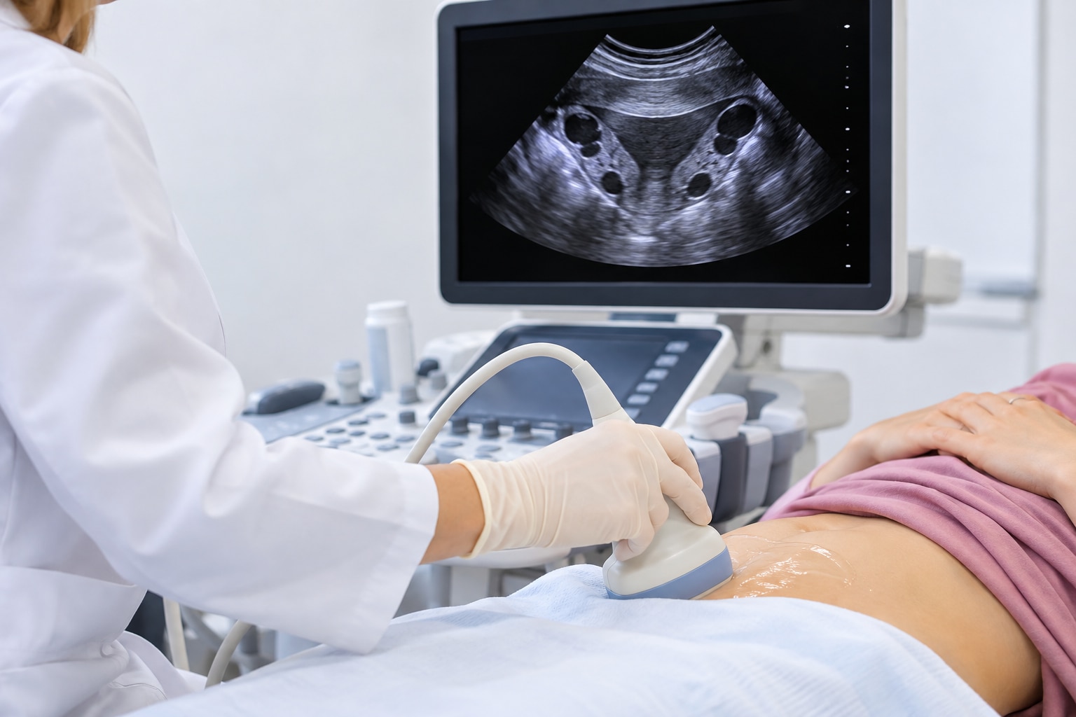
The choice depends on the clinical question, your symptoms, pregnancy status, and whether an internal examination is acceptable. A transabdominal pelvic ultrasound scans through the lower abdominal wall; a transvaginal ultrasound places the probe closer to the pelvic organs.
| Method | How it is performed | Strengths and limitations |
|---|---|---|
| Transabdominal | Gel is applied to the lower abdomen while you lie with a full bladder ultrasound | Provides a broader external view, but bowel gas or body habitus can reduce image quality. |
| Transvaginal | A covered, lubricated probe is gently placed in the vagina, usually after an empty bladder transvaginal scan preparation | The closer position gives higher-resolution views of the uterus, endometrium, ovaries, and adnexa, but it is an internal examination. |
The sonographer must explain the transvaginal ultrasound, answer your questions, and obtain your consent before starting. You may feel pressure or brief discomfort, and you should receive privacy and appropriate draping throughout the examination.
You can decline an internal scan, request a chaperone, or ask whether transabdominal imaging is an acceptable alternative. The external approach may provide less detail, so the best option depends on the structures that need assessment rather than on a fixed age group, pregnancy stage, or diagnosis.
When is pelvic ultrasound urgent, especially during pregnancy?
Routine referral is appropriate for stable symptoms, but pelvic pain in anyone who could be pregnant needs prompt assessment with a pregnancy test. A pelvic ultrasound pregnancy assessment can look for an intrauterine pregnancy, ectopic pregnancy, free fluid, or another cause of pain; an ectopic pregnancy ultrasound may not settle the diagnosis early.
Seek an emergency department or immediate medical care rather than waiting for a routine appointment if you have:
- Severe one-sided pelvic pain
- Fainting or near-fainting
- Shoulder-tip pain
- Marked weakness
- Heavy vaginal bleeding
A very early pregnancy or negative scan may not establish the diagnosis. Repeat pregnancy testing, serial blood tests, or follow-up ultrasound may be needed.
For acute pelvic pain, ultrasound with Doppler assesses the uterus, ovaries, adnexa, free fluid, and blood flow. Doppler flow alone does not exclude ovarian torsion; the symptoms and complete ultrasound appearance still matter.
For postmenopausal bleeding, transvaginal ultrasound is often an initial test. An endometrial thickness of 4 mm or less has greater than 99% negative predictive value for endometrial cancer in this setting, but persistent or recurrent bleeding still needs further evaluation, often including endometrial sampling.
Routine transvaginal screening for ovarian cancer is not recommended for average-risk, symptom-free women. Symptoms, a strong family history, or a hereditary cancer syndrome require an individual clinical plan rather than screening by ultrasound alone.
How should you prepare, and what happens during the appointment?
Pelvic ultrasound preparation depends on the method booked. Follow the centre’s instructions rather than assuming fasting is required. Do not fast unless the centre separately tells you to.
1. For a transabdominal scan, drink the instructed amount of water and arrive with a comfortably full bladder. It creates an acoustic window and moves bowel gas away from the pelvis. Avoid emptying your bladder until staff say it is acceptable.
2. For a transvaginal scan, empty your bladder first. You will usually change or lower clothing, lie on the examination bed, and receive privacy and draping. The covered, lubricated probe is inserted into the vagina only after the procedure is explained and you consent.
3. During the scan, the sonographer applies gel for external images and obtains views from several angles. You may be asked to change position or hold still. Pressure is possible, but tell the sonographer immediately if it hurts.
4. Menstruation or light spotting usually does not prevent examination. Ask about local instructions for ultrasound during menstruation or spotting before pelvic scan, and use a pad rather than a tampon at the appointment. Sexual activity does not determine whether the scan is appropriate.
Before imaging, mention pain, possible pregnancy, prior surgery, or discomfort with internal imaging. For what happens during transvaginal ultrasound, you can request an explanation, chaperone, pause, or alternative transabdominal approach.
What can the report show, and when is follow-up needed?
Pelvic ultrasound results describe what the scan can see, not every possible cause of symptoms. It may identify fibroids, adenomyosis, endometrial abnormalities, ovarian cysts, adnexal masses, pelvic inflammatory changes, and some endometriosis-related findings. A normal scan does not exclude superficial endometriosis or every cause of pelvic pain or abnormal bleeding.
Endometriosis ultrasound limitations matter when symptoms continue despite a normal examination.
A complex or indeterminate mass is not automatically cancer. The next step depends on its appearance, symptoms, age, and menopausal status:
- Follow-up ultrasound to monitor change
- MRI for further tissue characterisation
- Specialist review for clinical and imaging assessment
- Surgery when the appearance or symptoms require removal or diagnosis
For infertility, ultrasound can assess follicles, ovarian reserve markers such as antral follicles, and ovulation-related changes, but it cannot establish whether the fallopian tubes are open. Tubal patency requires a test such as hysterosalpingography or contrast-enhanced sonography.
An IUD position ultrasound usually locates a misplaced device; three-dimensional ultrasound can improve detection of subtle embedment when available.
When arranging a scan through Augmentum Diagnostics, provide:
- The referral reason and symptoms
- Your last menstrual period
- Any possibility of pregnancy
- Menopausal status
- Preparation instructions from the referring clinician
Those details match the appointment to the clinical question rather than treating it as generic screening.
Frequently asked questions
When do doctors recommend a female pelvic ultrasound?
Doctors may recommend it for pelvic pain, unusual bleeding, menstrual changes, fertility concerns, suspected fibroids or ovarian cysts, and abnormal examination findings.
Which is better: transabdominal or transvaginal ultrasound?
A transabdominal scan views the pelvis through the lower abdomen and usually requires a full bladder. A transvaginal scan uses a covered probe inside the vagina and gives closer images of the uterus, endometrium, and ovaries when appropriate.
When is a pelvic ultrasound urgent during pregnancy?
Seek urgent medical care for severe or one-sided abdominal pain, heavy bleeding, fainting, shoulder-tip pain, or pain with dizziness during pregnancy.
How should you prepare for a pelvic ultrasound?
Follow the centre’s instructions. A transabdominal scan often requires drinking water and keeping your bladder full, while a transvaginal scan usually requires an empty bladder.
What can a pelvic ultrasound report show?
The report can describe the uterus, endometrium, cervix, ovaries, adnexa, bladder, fluid, cysts, fibroids, masses, and pregnancy-related findings. Your clinician decides whether follow-up is needed.