

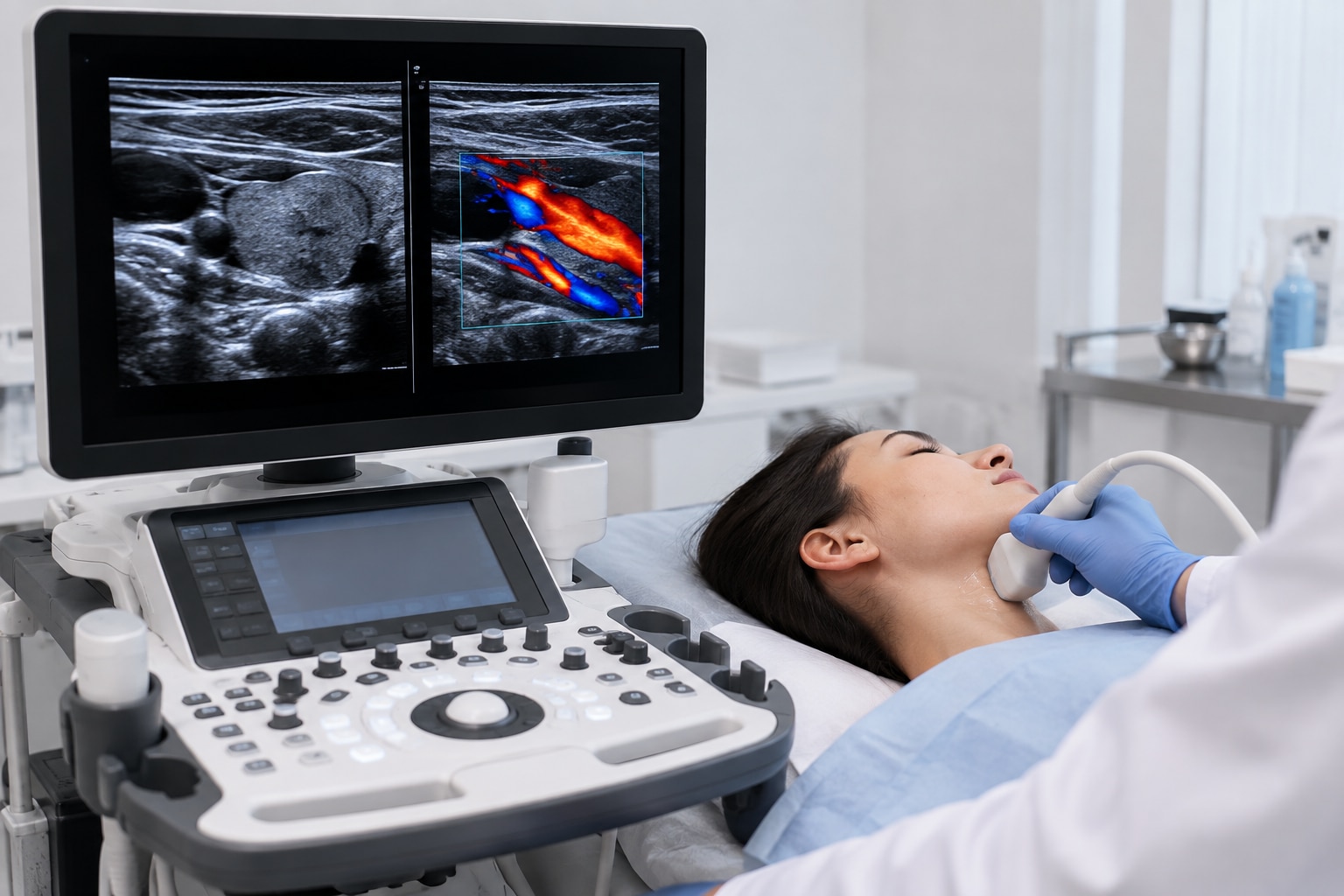
A standard ultrasound answers a structural question: what do the organs, tissues, fluid spaces, or developing pregnancy look like? Color Doppler adds a blood-flow question, showing movement within vessels or organs, so you can understand why a doctor requests one scan, the other, or both.
Key takeaways
- Standard ultrasound shows anatomy and tissue structure in grayscale.
- Color Doppler maps blood-flow direction and speed using color signals.
- Doctors request both scans when structure and circulation need assessment.
- Confirm the scan name, body area, preparation, and Doppler requirement before booking.
What a Standard Ultrasound Shows
Conventional ultrasound creates an image by sending high-frequency sound waves into the body through a handheld transducer. Different tissues and boundaries return echoes at different strengths and timings. A computer processes those echoes into B-mode imaging, producing the grayscale image seen on the screen.
This shows the size, shape, position, and texture of structures without ionizing radiation, unlike X-rays and CT scans.
A standard examination can assess:
- Abdominal organs such as the liver, gallbladder, pancreas, spleen, and kidneys, including findings such as gallstones, cysts, enlarged organs, or fluid
- Pelvic organs, including the uterus, ovaries, and bladder
- Thyroid, breast, testes, muscles, tendons, and other superficial soft tissues
- Developing pregnancies, including fetal growth, position, and visible anatomy
- Collections of fluid, enlarged lymph nodes, and many solid or cystic masses
Conventional ultrasound shows anatomy and tissue structure; it does not directly show how blood is moving. A grayscale scan can reveal a narrowed-looking vessel or a mass, but it cannot reliably establish blood-flow direction or speed. That question requires Color Doppler as an added mode or a targeted Doppler examination, depending on the requested protocol.
Because gas-filled bowel and bone block or weaken sound transmission, deeper targets and some body areas can produce incomplete or less-clear images.
What Color Doppler Adds to the Image
Color Doppler overlays colour on the grayscale ultrasound image to estimate blood-flow direction and relative flow speed. It uses Doppler frequency shifts from moving blood cells, assigning colour to sampled regions rather than replacing the structural image.
Red and blue usually indicate movement toward and away from the transducer, respectively. They do not mean artery versus vein, or oxygenated versus deoxygenated blood; the operator can reverse the colour map. The colour bar on the screen explains that examination’s direction and speed scale, while changes in shade usually represent different estimated velocities.
Color Doppler does not directly measure the total volume of blood passing through a vessel. Quantitative flow assessment also requires vessel diameter, the Doppler angle, the waveform, and assumptions about the flow pattern. An angle above 60 degrees creates substantially more velocity error.
A colour mosaic does not automatically prove reversed flow. Aliasing occurs when velocity exceeds the system’s Nyquist limit, producing a wraparound of colours; adjusting the velocity scale, pulse-repetition frequency, baseline, or Doppler frequency can reduce it.
The techniques answer different questions:
| Technique | What it displays | Main use |
|---|---|---|
| Color Doppler | Location, direction, and estimated relative speed | Find vessels or abnormal flow |
| Pulsed-wave spectral Doppler | Velocity waveform over time at a selected depth | Measure flow at a specific site |
| Continuous-wave Doppler | Very high velocities without exact depth localisation | Assess exceptionally fast flow |
Spectral Doppler is usually needed when velocity measurement matters.
When Doctors Request Ultrasound, Color Doppler, or Both
The suspected condition determines whether the examination needs anatomy, blood-flow information, or both. A routine ultrasound is appropriate when the main question concerns an organ’s size, shape, tissue pattern, fluid, or a mass. Color Doppler is added when movement of blood, vessel narrowing, blockage, reflux, or tissue perfusion could change the diagnosis.
- Request structural ultrasound for questions such as gallstones, kidney stones, liver lesions, thyroid nodules, or fluid collections.
- Request a targeted Color Doppler study when the concern is blood flow in the carotid arteries, venous reflux in varicose veins, organ perfusion, or flow around a lesion.
- Request both when anatomy and circulation must be judged together. Suspected testicular torsion needs grayscale images of the testicles plus Doppler comparison of their blood supply; a structural scan alone cannot reliably answer that question.
- For suspected deep-vein thrombosis, the examination uses vein-compression findings with appropriate venous Doppler imaging. Color Doppler alone is not a universal substitute for compression.
The body part also shapes the protocol. A carotid examination measures vessel structure and flow-related findings, while a venous leg study looks for compressibility, obstruction, and reflux. Tell the imaging centre the exact symptom and suspected diagnosis, because “ultrasound” and “ultrasound with Doppler” do not identify interchangeable protocols.
A Doppler color overlay complements, rather than replaces, the grayscale image. It estimates blood motion within the sampled area; it does not by itself measure total blood volume passing through the vessel.
What Happens During the Examination and What Can Limit It
Expect to lie on an examination bed while the sonographer exposes the area being scanned. They apply ultrasound gel and move an external probe across your skin, sometimes pressing firmly or asking you to change position, hold your breath, or keep a full bladder.
A transvaginal ultrasound uses a covered probe inserted into the vagina after you empty your bladder; the sonographer explains the procedure and obtains your consent.
Color Doppler usually takes place during the same appointment. The operator adjusts the probe and Doppler settings while viewing grayscale anatomy, so a flow finding is not interpreted from colour alone.
| Limitation | What goes wrong | Effect on the examination |
|---|---|---|
| Bowel gas | Traps and scatters the sound beam | Organs behind it become partly or completely hidden |
| Bone | Reflects or attenuates sound strongly | Structures behind ribs, the skull, or other bone are difficult to assess |
| Body habitus or deep target | The beam loses strength before reaching the target | Images contain less detail and Doppler signals weaken |
| Motion | Patient, breathing, or probe movement disrupts sampling | False colour, blurred anatomy, or unreliable measurements |
| Doppler settings | Poor angle, excessive wall filter, wrong gain or frequency, or aliasing distorts flow display | A weak or absent colour patch can be mistaken for absent blood flow |
Doppler cannot overcome these physical barriers. The operator must match the technique to the question: suspected deep-vein thrombosis requires vein compression assessment with suitable venous Doppler imaging, not color Doppler alone. A limited view or uncertain signal belongs in the report and may require another approach.
How to Confirm the Correct Scan Before You Book
Before you book, make sure the order names the body part, side, clinical question, and required technique. An ultrasound referral for a lump, abdominal pain, pregnancy, or suspected vessel problem does not automatically mean the same examination.
1. Confirm the exact examination name. Ask whether the doctor wants routine grayscale imaging, a targeted Doppler study, or both. For suspected deep-vein thrombosis, confirm that venous compression assessment is included; Color Doppler alone is not a complete substitute.
2. Confirm the location and coverage. The order should identify the organ, vessel, limb, or lesion, plus the right or left side when relevant. Ask whether the scan must assess blood flow, narrowing, blockage, organ perfusion, a mass’s vascularity, or pregnancy blood flow.
3. Give the centre the clinical details and previous results. Provide symptoms, their duration, pregnancy status and gestational age when relevant, surgery history, and earlier scans. These details help the sonographer select the scan protocol and interpret an absent or reduced color signal correctly.
4. Ask about preparation and timing. Fasting, a full bladder, medication instructions, or urgent scheduling can affect the examination. In early pregnancy, ask why Doppler is needed and confirm that exposure will remain as low and brief as reasonably possible.
Augmentum Diagnostics can clarify whether its booking entry matches the written referral before you attend. Check that the final diagnostic report will name the structures examined, Doppler measurements when required, technical limitations, and the clinical question it addresses.
Frequently asked questions
What does a standard ultrasound show?
A standard ultrasound produces grayscale images of organs, tissues, fluid, and anatomical boundaries using reflected sound waves.
What does Color Doppler add to an ultrasound?
Color Doppler adds a visual map of moving blood, helping assess flow direction and relative speed in vessels.
When do doctors request ultrasound, Color Doppler, or both?
Doctors use standard ultrasound to assess anatomy, Color Doppler to assess blood flow, and both when structure and circulation matter.
What happens during an ultrasound or Color Doppler examination?
You usually lie down while a sonographer applies gel and moves a transducer over the examination area; preparation and duration depend on the scan.
What can limit an ultrasound or Color Doppler examination?
Gas, body tissue thickness, movement, limited access, and an unfilled or overly filled bladder can reduce image quality.