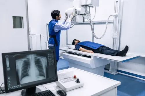


Abdominal discomfort can arise from many different organs and conditions. When symptoms cannot be explained through clinical examination alone, a doctor may recommend imaging at an Ultrasound Center in Mahavir Enclave to obtain additional information about structures inside the abdomen.
Ultrasound is widely used because it creates real-time images using sound waves rather than ionising radiation.
What Can an Abdominal Ultrasound Examine?
Depending on the investigation requested, ultrasound may help assess structures such as the:
- Liver
- Gallbladder
- Kidneys
- Spleen
- Urinary bladder
- Pancreas where adequately visualised
- Selected abdominal structures
The exact examination depends on the patient's symptoms and the clinical question being investigated.
Symptoms That May Lead to an Ultrasound
An ultrasound is not required for every episode of stomach discomfort. A doctor may consider imaging when symptoms persist, recur, or suggest that an internal organ should be evaluated.
Possible reasons include persistent abdominal pain, bloating associated with other symptoms, urinary concerns, suspected gallbladder problems, unexplained abdominal findings, or abnormal laboratory results.
The decision should be based on clinical evaluation rather than simply undergoing scans whenever discomfort occurs.
Why Ultrasound Is Commonly Used
One major advantage is that ultrasound does not use ionising radiation.
It can also provide real-time images, making it useful for evaluating several abdominal and pelvic structures.
However, ultrasound has limitations. Image quality can be affected by bowel gas, body characteristics, patient preparation, and the anatomical area being examined.
For some medical questions, another imaging technique may be more appropriate.
Preparation Can Affect the Examination
Patients should pay close attention to instructions provided before their appointment.
Some abdominal ultrasound examinations may require fasting. Other examinations involving the urinary bladder or pelvic structures may require a comfortably full bladder.
Preparation requirements vary according to the type of scan.
Following these instructions helps the sonologist or radiologist obtain useful images and may reduce the likelihood of an incomplete examination.
What Happens During the Scan?
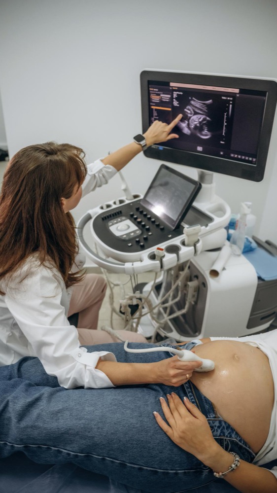
The patient generally lies on an examination couch while ultrasound gel is applied to the relevant area. A handheld transducer is then moved over the skin.
The transducer sends sound waves into the body and receives returning echoes. These signals are processed into images that can be evaluated.
The examination is generally non-invasive, although pressure from the transducer may cause temporary discomfort if the area is already tender.
Why Professional Interpretation Matters
Producing an image is only one part of diagnostic imaging.
The findings must also be evaluated in relation to anatomy and the clinical question. Augmentum Diagnostics provides ultrasound services as part of its diagnostic offering, with the objective of producing clinically useful reports for further medical evaluation.
Patients should remember that imaging findings are usually one part of a larger diagnostic picture.
A doctor may consider the ultrasound alongside physical examination, blood tests, urine tests, previous imaging, and medical history.
Ultrasound Does Not Replace Medical Consultation
It can be tempting to arrange an ultrasound independently when abdominal symptoms continue. However, abdominal pain can have many causes, and not every cause can be diagnosed with ultrasound.
Severe or rapidly worsening pain, persistent vomiting, fainting, blood in stool or vomit, significant breathing difficulty, or other alarming symptoms require prompt medical assessment.
For appropriately selected cases, an Ultrasound Center in Mahavir Enclave can provide valuable information without exposing the patient to ionising radiation. The most useful results come from combining proper preparation, appropriate imaging, professional interpretation, and clinical follow-up.